



Exosomes Explained: Inside Skincare’s Tiniest Delivery System
Skincare's smallest delivery system, explained — how exosomes are taken up by skin cells, and what our Cica exosomes were clinically proven to do.
Author
askINKEY skincare advisor
Published
16 January, 2026
Time to read
20 minutes
Exosomes are nano-sized vesicles measuring 30 to 150 nanometres across. Each one is enclosed by a lipid shell and carries a cargo of lipids, proteins and genetic material inside it. Skin cells take them up through three recognised routes: direct membrane fusion, endocytosis, and receptor docking. To give that size some context, an exosome is around 300 times smaller than a pore.
That is the short answer to how do exosomes work. The rest of this article is the long one.
Most exosome content online tells you two things. It tells you exosomes are very small, and it tells you they are a breakthrough. Then it stops. What is missing almost everywhere is the actual physical journey: what an exosome is built from, what is held inside it, how it was packed and sealed before it ever reached the bottle, and what happens at the moment it meets a skin cell. That journey is the interesting part, and it is also the part that separates genuine understanding from marketing language you cannot verify.
So this article does one job properly rather than several jobs loosely. It covers structure, cargo and absorption. It explains how are exosomes absorbed in real mechanical terms, using the peer-reviewed literature as the reference point rather than metaphor alone. It is a delivery story, and it follows the parcel from the bottle to the doorstep.
What it deliberately does not cover: which benefits to expect, where exosomes sit in your routine, how they compare to other actives, or which product suits your skin type. Those are decision questions rather than mechanism questions, and they are answered properly in our complete exosome guide. If you came here to choose a product, go there instead. If you came here because you want to know what is actually happening when the serum lands on your face, stay.
We are going to follow one exosome from bottle to skin cell. The journey starts with the thing that makes all of it possible: its size.
How Small Is An Exosome? Understanding Nanometre Scale
Numbers in skincare are usually there to impress rather than inform. This one is different, because the size of an exosome is not a selling point bolted onto the science. It is the precondition for the science working at all.
Exosomes measure 30 to 150 nanometres in diameter. A nanometre is one billionth of a metre. That phrase is easy to read and almost impossible to picture, so it is worth building a comparison ladder before going any further.
Start at the top of the ladder with something you can see. A single human hair is roughly 80,000 to 100,000 nanometres wide. Drop down a rung: a red blood cell, invisible to the naked eye, measures around 7,000 nanometres. Drop down again to a pore on your face, which sits at around 50,000 nanometres across. Now drop all the way to the bottom rung. An exosome is 30 to 150 nanometres.
Read those figures next to each other and the gap is genuinely difficult to hold in your head. A pore is not slightly larger than an exosome. It is enormously, structurally larger. Which brings us to the anchor figure worth remembering: the Cica exosomes in our formula are around 300 times smaller than a pore, and there are 3 million plant-derived exosomes in every bottle.
Why an exosome is not just another small ingredient
Here is where scale stops being a party trick and starts being meaningful.
Almost every active ingredient in your bathroom cabinet is a single molecule. Vitamin C is a molecule. Niacinamide is a molecule. Salicylic acid is a molecule. They are individual chemical units, and their behaviour on skin is governed by their own molecular properties: size, charge, solubility, stability.
An exosome is not a molecule. It is a fully enclosed structure. It has an outer shell and it has contents held inside that shell, which makes it closer in architecture to a container than to a chemical. That distinction matters more than the size figure on its own.
Being enclosed is not unusual. Being nano-sized is not unusual. Being both at once is where exosomes become genuinely interesting, because it means you have a sealed container operating at a scale where sealed containers do not normally exist in skincare formulation. The shell holds the cargo together. The size lets the whole assembly reach the right neighbourhood on skin.
Small alone is not enough, and we should say so
There is a version of this article that would stop here, declare that small equals effective, and move on to the product. That version would be dishonest.
Size gets a particle to the right place. It does not determine what happens when it arrives. Plenty of small things sit on the surface of skin and do very little, because reaching a location is not the same as being accepted at that location. A parcel arriving at your front door has completed only part of its journey. Whether it comes inside depends entirely on what it looks like at the door.
What happens next depends on what the particle is made of. That is the next section, and it is the more important of the two.
Does topical absorption require microneedling?
This question comes up constantly, so it is worth answering here rather than burying it, because it is genuinely a mechanism query rather than a product one.
No. Topically applied plant-derived exosomes at this scale are absorbed without needling. The reason sits in the numbers above. Microneedling exists to create physical channels through the skin’s outer layers, which is relevant for larger molecules that struggle to get anywhere without help. At 30 to 150 nanometres, enclosed in a shell that is chemically compatible with the environment it is landing in, an exosome is not facing that problem. The channel-making step is solving an obstacle that does not apply at this scale.
We will return to the practical and financial side of that comparison later on. For now, the mechanical point stands on its own: needling is not a requirement for topical absorption.
Size explains how an exosome reaches the surface of your skin. It does not explain why your skin accepts it once it arrives. That comes down to what the outer shell is built from.
The Lipid Shell: Why Skin Recognises An Exosome
Every exosome is wrapped in a phospholipid bilayer. That term deserves unpacking rather than skipping past, because it is the single most important structural fact in this entire article.
A phospholipid is a fat molecule with two ends that behave differently: one end is drawn to water, the other end avoids it. Put a very large number of them together in a watery environment and they arrange themselves automatically into a double-layered sheet, with the water-avoiding ends tucked inward, facing each other, and the water-loving ends facing outward on both sides. That double-layered sheet is a bilayer. Curve it around into a closed sphere and you have an enclosure.
Now the part that matters. This is not a specialist structure invented for exosomes. It is the same fundamental architecture used by cell membranes throughout living things. Your own skin cells are wrapped in a phospholipid bilayer. Plant cells are wrapped in one. It is the standard building material of biological containers, which means an exosome’s outer shell and a skin cell’s outer membrane are built from the same class of material.
The shell around an exosome is not a coating applied to it. It is made from the same class of material as the membranes of your own skin cells, which is why the two are chemically compatible in the first place.
Like dissolves like, applied to skin
There is an old principle in chemistry: like dissolves like. Oil-based substances mix readily with oil-based substances. Water-based substances mix readily with water-based substances. Try to combine across that divide and things separate.
The outer layers of your skin are lipid-rich. The spaces between the cells of the stratum corneum are filled with a mortar of lipids, including ceramides, cholesterol and fatty acids. It is a fatty, oily environment by design.
A lipid-shelled particle arriving in a lipid-rich environment is therefore chemically at home. There is no fundamental incompatibility to overcome, no mismatch of character between the visitor and the location. This is the actual reason exosome absorption is plausible, and it is considerably more convincing than any marketing sentence about penetration, because it rests on the composition of the particle rather than on an assertion about performance.
It is worth pausing on how unusual that is. Most delivery challenges in skincare formulation involve persuading an ingredient to be somewhere it is not naturally suited to being. Exosomes arrive already suited.
The shell as protective packaging
The bilayer is doing a second job at the same time, and it is easy to overlook.
An enclosure protects what is inside it. The cargo held within an exosome is not floating loose in your serum, exposed to everything around it. It is held inside a sealed container while the particle is in transit. That is why the contents are still intact by the time the particle reaches skin cells rather than degraded somewhere along the way.
Think of it as the difference between posting a delicate object loose in a sack and posting it boxed. The box is not the point of the delivery, but without the box there is nothing worth delivering by the time it arrives.
Surface proteins: the label on the parcel
Sitting within the lipid shell are proteins that stud its surface. Among the most consistently identified across the research literature are a family called the tetraspanins, including CD9, CD63 and CD81.
It is important to be precise about what these are, because this is exactly where a lot of exosome content overreaches.
Surface proteins function as identification and docking compatibility. They are the label on the parcel and the shape of the connector on the end of the cable. A label tells you what the parcel is. A connector either fits the socket or it does not. Neither the label nor the connector commands anything, and neither one is an instruction.
That is the whole of it. These proteins determine whether a given exosome and a given cell surface are physically compatible with one another. They govern fit, not consequence. Any description that goes further than fit is going further than the structural biology supports.
This is structural biology, not a formulation trick
Everything described in this section is a feature of what an exosome fundamentally is. The bilayer is not a delivery technology wrapped around an ingredient in a lab. The surface proteins are not added during manufacture. This is the native architecture of the particle, present from the moment it was formed inside its source cell.
Our Cica exosomes are plant-derived, sourced from Centella Asiatica, and they arrive already enclosed in exactly this shell. If you want to go deeper on where exosomes come from and how sourcing shapes what you end up with, we have written separately about plant-derived versus fermentation-derived exosomes.
One caveat before we move inward. A shell this fine needs a supportive formula around it, one built to keep the enclosure intact rather than compromise it. Ectoin plays a part in that, and we will come back to it properly when we get to formulation.
We have now covered the outside of the particle. The obvious question is what is actually inside it.
What Exosomes Carry: Lipids, Proteins And Genetic Material
Open the parcel. What is in there?
Exosome cargo falls into three broad classes, and each one is worth defining in plain terms rather than leaving as jargon.
Lipids. Fats. Not the ones in your diet, but the structural kind: the molecules that make up and support membrane structures. Lipids appear both in the shell itself and among the contents held inside it.
Proteins. The functional building blocks of biology. This category is broad and includes peptides, which are short chains of amino acids, and enzymes, which are proteins with specific chemical roles. If you want the background on how peptides work as a skincare ingredient class in their own right, our guide to peptides covers it, and it is relevant here because Kollaren peptide sits in the hero formula alongside the exosomes.
Genetic material. Information-carrying molecules, principally RNA, held within the vesicle. This is the cargo class that generates the most excitement and, frankly, the most overstatement in consumer content. What can be said accurately is that these molecules are present inside the vesicle and are carried by it.
Cargo is selected, not random
This is the detail that most articles miss, and it changes how you should read any exosome claim.
Vesicles are not filled arbitrarily. Different source cells produce vesicles carrying different contents, and those contents vary depending on the conditions the source cells were grown in. Cargo profile is a direct function of source.
The practical consequence is significant. “Exosomes” is not a single ingredient in the way that “niacinamide” is a single ingredient. It is a category of structure. Two exosome products can both accurately be called exosome products and contain vesicles with meaningfully different contents inside them, because they came from different sources grown under different conditions.
Anyone who tells you all exosomes are equivalent is either simplifying heavily or has not thought about it. Anyone who tells you exosomes do a specific thing without telling you where those exosomes came from has left out the variable that determines the answer.
What Cica exosomes carry
Our exosomes are derived from Centella Asiatica, which means the vesicles carry a cargo profile characteristic of that plant. Centella Asiatica has a long and well-documented history in soothing skincare preparations, and the exosomes derived from it reflect their source, as all exosomes do.
Two measured findings are associated with the ingredient. Both come from in-vitro testing, meaning laboratory testing rather than testing on people, and both are reported here exactly as they were measured:
55% reduction in visible signs of skin stress (redness, puffiness, visible irritation)***
63% increase in skin renewal activity in 8 hours***
Those are outcomes associated with the ingredient, and they sit separately from the mechanism description in the rest of this article. The mechanism story is about how a sealed container reaches a cell and releases what it holds. The measured findings are a different kind of evidence, generated in a different setting, and it would be sloppy to blur the two together.
Being honest about the limits
Plant-derived exosome research is active and developing. That sentence is not a hedge, it is the accurate state of the field.
What is thoroughly described in the literature is the structural side. The size range, the bilayer architecture, the surface protein families, the packing pathway, the uptake routes: this is established cell biology, replicated across many studies, and it is what this article is built on.
What is still being worked out is the application side, particularly for plant-derived vesicles specifically, which are a newer area of study than their mammalian counterparts. There is a great deal of research happening and a great deal still to establish.
We would rather tell you that than pretend the whole field is settled. Overclaiming is how ingredient categories get burned, and exosomes are genuinely interesting enough that they do not need the help. If you have been reading exosome content elsewhere and found yourself unsure what to believe, we have gone through the most common misconceptions in 5 exosome skincare myths debunked. The composition and behaviour of plant-derived nanovesicles is also covered in depth in the peer-reviewed literature on plant-derived exosome-like nanoparticles for anyone who wants to read the primary sources.
We know what is in the parcel. The next question is how it got in there, and how it was sealed. For that, we have to go inside the source cell.
How Exosomes Are Packed And Sealed: Biogenesis And The ESCRT Pathway
This is the part almost nobody explains, and it is the part that makes everything else make sense.
Exosomes are not assembled at the surface of a cell and pushed out. They are built deep inside the source cell, packed and sealed there, and released fully formed. There is a packing line, it has stages, and it has dedicated machinery. Understanding it tells you something practical: by the time an exosome is in a bottle, the sealing already happened a long time ago, somewhere else.
Let us walk the line.
The endosomal pathway, step by step
Step one: the membrane folds inward. The outer membrane of the source cell dips inward, forming a small pocket that pinches off into the interior of the cell. That newly formed internal compartment is called an early endosome. Picture the surface of a balloon being pushed in with a fingertip until the indentation separates and floats free inside.
Step two: the endosome matures. The early endosome develops into a late endosome. This is a maturation process, with the compartment changing in composition and character as it moves inward through the cell.
Step three: vesicles bud inward, inside the compartment. Now the same inward-folding motion happens again, but this time on the membrane of the late endosome itself, and it happens repeatedly. Small vesicles bud inward and accumulate inside the larger compartment. You end up with a container full of smaller containers.
Step four: the multivesicular body. That structure, a compartment holding many small vesicles inside it, has a name: a multivesicular body. The small vesicles held within it are called intraluminal vesicles, meaning vesicles inside the lumen, or interior space.
Step five: release. The multivesicular body travels to the cell’s outer membrane, meets it, and opens. The intraluminal vesicles held inside are released out of the cell.
Those released vesicles are exosomes. Same structures, new name, determined by location. Inside the multivesicular body they are intraluminal vesicles. Once released, they are exosomes.
ESCRT: the packing and sealing machinery
Something has to do the packing. That something is a set of protein complexes with an unwieldy name given in full once here: Endosomal Sorting Complexes Required for Transport, universally shortened to ESCRT. The machinery operates in four stages, supported by a recycling component, and it functions very much like an assembly line.
-
ESCRT-0 gathers the cargo. The first complex collects and clusters together the material that is going to be packed, bringing it into one place on the membrane rather than leaving it scattered.
-
ESCRT-I and ESCRT-II draw the membrane inward. With the cargo clustered, these complexes begin pulling the membrane in around it, forming a developing pocket with the cargo held inside. The pocket deepens, and a narrow neck forms behind it where it remains connected to the parent membrane.
-
ESCRT-III pinches and severs the neck. The third complex assembles at that narrow neck, constricts it, and cuts through. The pocket closes fully and separates. The vesicle is now sealed, with its contents enclosed inside.
-
VPS4 dismantles the machinery. A separate component breaks the ESCRT assembly apart afterwards so the components can be reused elsewhere. Nothing is wasted, and the line resets for the next parcel.
Read that sequence back and notice what all the verbs have in common. Gathers, clusters, draws inward, pinches, severs, closes, seals, dismantles. Every one of them is structural. This is a physical packing operation, and describing it as anything more than that would be adding a layer the biology does not contain.
The ceramide route, and a skincare connection
ESCRT is the best-characterised packing route, but it is not the only one. Alternative, ESCRT-independent pathways also exist, and one of the most studied involves ceramides.
Ceramides are lipids, and they have a distinctive cone-like molecular shape. Where ceramides accumulate in a patch of membrane, that shape encourages the membrane to curve. Enough curvature in one place and the membrane buds inward on its own, forming a vesicle without the protein machinery driving it. Geometry does the work instead.
If ceramides sound familiar, that is because you almost certainly already own some. They are a cornerstone skincare ingredient, and the same lipids that support the skin’s outer layers are involved in membrane curvature at the cellular level. It is a genuine crossover between cell biology and the ingredient list on your bathroom shelf rather than a stretched analogy. Our ceramides guide covers their role in skincare properly, and they are the basis of our Bio-Active Ceramide Moisturiser, which we will come back to at the end.
Why any of this matters to you
Here is the payoff, and it is worth stating plainly.
All of this packing and sealing happens inside the source plant cells, long before anything reaches a laboratory, let alone a bottle. Which means the exosomes in a serum arrive pre-packed and already sealed.
Nothing is assembled on your face. No packing step is happening in the bottle or on your skin. The vesicle you apply was built, filled and closed by the plant cell it came from, and it has been an intact sealed container ever since. That is precisely why the cargo is still enclosed and intact when it reaches skin, and it is the reason the whole delivery premise holds together rather than being an aspiration.
It also explains why sourcing is not a marketing detail. The packing line that built your exosomes was inside a specific plant, grown under specific conditions, and that determined what got packed. The parcel was addressed and filled before it ever entered the supply chain.
The parcel is packed, sealed and released. Which leaves the question most people are actually searching for: how does it get into a skin cell?
How Are Exosomes Absorbed? Membrane Fusion, Endocytosis And Receptor Docking
There is no single answer, and any article that gives you one is oversimplifying.
Skin cells take up exosomes by at least three recognised mechanisms, and which one occurs in a given instance depends on the cell involved and the conditions at the time. They are not competing theories where one will eventually be proven correct. They all happen. Understanding how exosomes are absorbed means understanding all three.
Route one: direct membrane fusion
The fastest route, and the most elegant.
Remember that the exosome’s outer shell and the cell’s outer membrane are built from the same class of material. Two lipid bilayers, same fundamental architecture. When they come into contact under the right conditions, they can merge into one continuous membrane. The boundary between them stops existing.
At the moment of merging, the cargo that was enclosed inside the exosome is released directly into the interior of the cell. There is no intermediate compartment and no unpacking stage. The container becomes part of the cell wall, and what it held is now inside.
The analogy that fits best is two soap bubbles touching. They do not bounce off each other and they do not sit side by side. They merge, and what was inside both is now inside one.
This route is only possible because of the compatibility described earlier. It is a direct consequence of the shell being made of what it is made of, which is why the lipid section came before this one.
Route two: endocytosis
The most common route, and the one that accounts for the bulk of uptake.
Rather than merging with the exosome, the cell membrane curves around it. The membrane forms a depression, the depression deepens around the particle, the edges come together and close, and the exosome ends up enclosed inside an internal compartment within the cell. The whole parcel is taken inside intact rather than being opened at the boundary.
If that motion sounds familiar, it should. It is essentially the same inward-folding movement described in the biogenesis section, running in the same direction. Cells use this manoeuvre constantly.
Endocytosis is not one process but a family of related ones, and the recognised sub-routes are worth naming:
- Clathrin-mediated endocytosis, in which a protein called clathrin assembles into a scaffold on the inner face of the membrane, forming a cage that shapes the pocket as it draws inward.
- Macropinocytosis, a less selective route in which the membrane ruffles outward and folds back on itself, sweeping in a larger volume of surrounding fluid along with whatever it contains.
- Phagocytosis, in which the membrane extends around a particle and engulfs it, a mechanism most associated with specialised cells.
The analogy here is a parcel being taken inside the building and opened in a sorting room rather than at the doorstep. The delivery is completed, but the unpacking happens internally.
Route three: receptor docking
The most selective route, and the one where surface compatibility becomes decisive.
Surface proteins on the exosome and complementary proteins on the cell surface fit together and bind. This is a physical matching event: a connector meeting a socket that accepts its shape. If the shapes are compatible, they bind. If they are not, they do not, and the particle continues on its way.
That is the entire description, and it is deliberately where the description stops. Research into extracellular vesicle uptake covers both endocytic and non-endocytic routes including receptor engagement, and the well-established part is the binding itself: the fit, the compatibility, the physical connection between two surfaces.
Why not every cell takes up every exosome
The selectivity point deserves its own moment, because it is where the three routes tie together into something meaningful.
Uptake depends on surface compatibility. Not every cell will take up every exosome, because the surfaces have to be compatible for binding and, in the fusion case, for merging. Different cell types present different surface protein profiles. Different exosomes carry different surface protein profiles, determined by their source.
This is why source matters so much, and why “it contains exosomes” is an incomplete statement about a product. The source determined the cargo. The source also determined the surface profile, and the surface profile influences which cells are receptive.
It is also worth noting that different renewal ingredients reach skin by entirely different routes. Exosomes are taken up by skin cells as enclosed vesicles. Other ingredients arrive as free molecules and behave accordingly. PDRN is a good example of a completely different structural approach, and we have set the two side by side in PDRN versus exosomesfor anyone who wants the mechanical comparison.
Absorption depends on the exosome surviving the trip
All three routes share one requirement: the shell has to still be intact when it reaches skin.
A compromised shell is not a delivery vehicle. If the enclosure has broken down in the bottle, the cargo is no longer enclosed and there is nothing to be taken up as a unit. Fusion needs an intact bilayer to fuse with. Endocytosis needs a particle to enclose. Receptor docking needs surface proteins sitting in a stable membrane.
Which makes the surrounding formula part of the delivery system rather than a neutral background. This is also why exosome serums are applied first in a routine, straight after cleansing, so nothing sits between the formula and the skin obstructing absorption. We have written about how that plays out alongside other actives in boost your retinol results with exosome, and the complete exosome guide handles routine placement in full.
From the finished formula, the experience is more straightforward. Our exosome serum is Proven to deliver up to 12-hours of hydration**, which is the part of all this you can feel rather than the part you have to take on trust.
Three routes, one shared requirement. That requirement is a formulation question as much as a biological one.
From Biology To Bottle: How Formulation Protects Absorption
An exosome only works if it arrives intact. Everything in the previous five sections depends on that single condition.
Which means the formula around the exosome is doing real work. A heavy, occlusive or aggressive base can compromise a delicate enclosure. Get the surrounding formula wrong and you have an ingredient list that mentions exosomes and a product that cannot deliver them. The formula is part of the delivery system, not a carrier that happens to be there.
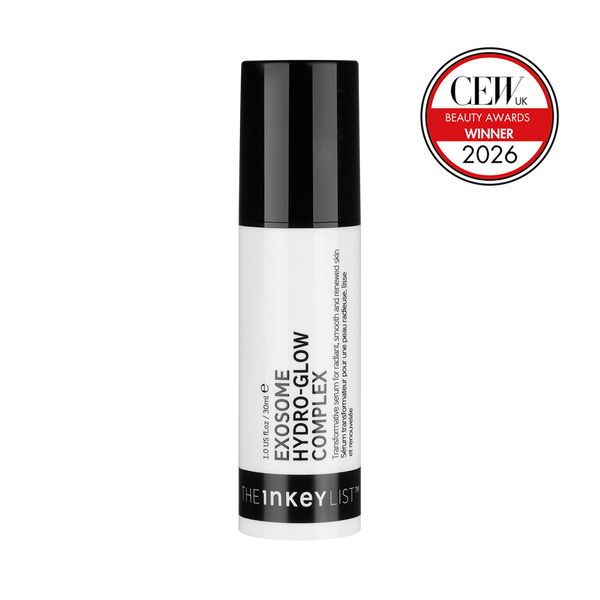
That is the thinking behind the Exosome Hydro-Glow Complex, £20.00 for 30ml, built around 1% plant-derived Cica exosomes from Centella Asiatica.
What each supporting active is doing
The other ingredients are not there to lengthen the list. Each one has a job connected to keeping the delivery environment right and the skin comfortable while it happens.
Hyaluronic Acid provides multi-level hydration, holding water at different depths within the skin’s upper layers rather than at a single level. Hydrated skin is a better environment for a lipid-shelled particle to arrive into, and hydration is the most immediate thing most people notice. If dryness and tightness are your main concern, our guide to dehydrated skin explains what is actually happening and why hydration behaves differently from oil.
Ectoin supports moisture retention and barrier support. It is a naturally occurring molecule that helps skin hold onto water and stay comfortable, and it is particularly useful in a formula where you want a calm, stable surface rather than a stressed one. There is more on how it works in our Ectoin guide.
Kollaren peptide supports firmness and skin structure. Peptides are short chains of amino acids, and this one is included for its role in the structural side of the formula.
Q10 provides antioxidant defence. Coenzyme Q10 is found naturally in skin and helps protect against environmental stressors.
Prickly Pear extract offers gentle resurfacing, contributing to smoothness without the harshness that would compromise a delicate delivery environment.
The texture follows the same logic. It is lightweight and creamy with no stickiness, and it is applied to damp skin straight after cleansing, morning and evening. Damp skin and a light texture both serve absorption rather than getting in its way.
On suitability: the formula is suitable for all skin types including sensitive skin, and it is safe to use during pregnancy and breastfeeding. Those are not incidental footnotes. For a lot of people they are the difference between a product being usable and being off the table entirely.
What the testing found
Across clinical testing, 100% saw more glowing skin, clinically proven*.
On timeframe, the study result was Clinic worthy rejuvenated skin in 14 days*.
Looking across the range of measures assessed, it delivers 6-in-1 skin rejuvenation: clinically proven to visibly improve radiance, hydration, tone, firmness, elasticity, and texture*.
The formula also Helps support natural collagen production.
Those are the findings as they were recorded. We have not rounded them, reframed them or extrapolated beyond them, and the study details sit in the footnotes at the bottom of this article so you can see exactly what was tested and on how many people. That is the standard we think ingredient claims should be held to, ours included.
The accessibility point
There is a cost conversation attached to exosomes that is worth having openly, because it shapes who gets access to this science.
In-clinic exosome treatments in the UK typically start from around £200 to £400 per session for topical protocols. Where treatment is combined with microneedling, that rises to roughly £350 to £600 per session. Clinics commonly recommend a course of three sessions rather than a single visit, which puts a full course somewhere between several hundred and well over a thousand pounds.
Set that against £20.00 for a 30ml bottle.
We are not going to pretend a serum and a clinical protocol are the same thing, because they are not. They are different formats delivered in different settings. But the underlying science, the nano-sized vesicle with its lipid shell and its enclosed cargo, is the same science. Making that available at £20.00 rather than at several hundred pounds a session is not a marketing angle for us, it is the reason the brand exists. We have written more about that thinking in the glow revolution.
The step that follows
Once the serum is on, a moisturiser seals it in. Our Bio-Active Ceramide Moisturiser at £20.00 is the natural companion, and it connects directly back to the ceramide science raised earlier. The same class of lipid involved in membrane curvature inside cells is the class of lipid supporting the outer layers of your skin.
If you want the full product story rather than the mechanism behind it, we have covered that separately in meet Exosome Hydro-Glow Complex.
The most common remaining questions about exosome absorption are answered directly below.
Conclusion: Small Particle, Clear Science
Here is the whole journey in one paragraph. An exosome is 30 to 150 nanometres across, around 300 times smaller than a pore. It is enclosed in a lipid shell built from the same class of material as your own cell membranes, which is why skin is chemically compatible with it. Inside that shell it carries lipids, proteins and genetic material, a cargo determined entirely by the source it came from. It was packed and sealed by ESCRT machinery deep inside a source plant cell, long before it reached the bottle, which is why it arrives intact. And skin takes it up through fusion, endocytosis or receptor docking, depending on the cell and the conditions.
That is a delivery system, described end to end, without a single step that requires you to take our word for it.
The honest closing point is this. Exosome science is thoroughly described at the structural level and still developing at the application level, particularly for plant-derived vesicles. We would rather tell you where the confident ground ends than blur the two together, because that distinction is the difference between education and hype. An ingredient this interesting does not need to be oversold.
What we will say plainly is that this level of science sitting in a £20.00 bottle is the entire point. Not a diluted version of something clinical, but the same structural biology, made accessible.
Skincare has become genuinely complicated, and most of that complication is not doing anything for you. Clarity you can actually trust is what helps. If you now want the benefits, the routine placement and the product guidance, that all lives in our complete exosome guide.
Because at The INKEY List, We Give A Care.
Ready To Put The Science On Your Skin?
If you have read this far, you know exactly what you would be putting on your face and why it works the way it does.
Shop the Exosome Hydro-Glow Complex, £20.00 for 30ml. Apply it to damp skin, straight after cleansing, morning and evening, so nothing gets in the way of absorption.
Want benefits, routine placement and guidance on whether it suits your skin? That is all in the complete exosome guide.
Related reading:
4-week clinical study of 26 people.
Clinical study of 31 people. In-vitro testing of Cica Exosomes showed a 55% reduction in pro-inflammatory markers and a 63% increase in markers associated with skin renewal after 8 hours.
Frequently Asked Questions
Got a question?
Our askINKEY team are a friendly bunch of skincare experts with deep product knowledge and zero judgement. Whether you're an ingredient obsessive or just starting out, we'll make it make sense. No question too big. No question too small. We Give A Care, Every Day.


